Why this review still sits in the SMART reference list
In January 1991, Mats Hanson and Jaro Pleva published The dental amalgam issue. A review in Experientia (47(1):9–22). IAOMT lists it as SMART Reference #8. It is an interdisciplinary review of materials science, corrosion, mercury exposure, toxicology, neurology, and immunology—plus new in-vivo measurements of mercury leaving corroded fillings and of mercury-vapour absorption inside the mouth.
Their conclusion is direct: mercury exposure from corroded amalgam can reach levels considerably over known threshold limit values. A galvanic amalgam–gold couple is not cosmetic; they call avoiding it of “vital importance.” They include a disabled patient who recovered after amalgam removal, and they close with the acrodynia lesson: when a neurological or immunological disease has no clear cause, amalgam mercury belongs on the differential.
Amalgam is not an inert “silver” filling
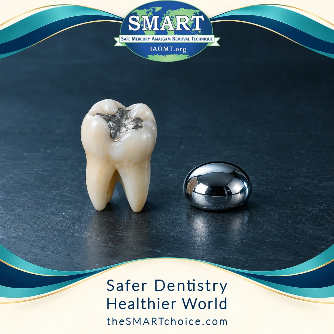
Hanson and Pleva argue that 150 years of amalgam debate failed because dentistry treated the filling as a mechanical object and medicine never saw the mercury source. The “silver filling” label withholds the essential fact: conventional silver amalgam is typically 50% mercury by weight (about 35% silver, 10% tin, plus copper and zinc). High-copper “non-gamma-2” products, introduced to blunt gamma-2 corrosion, simply shifted the corrosion-prone phases. The filling is still half mercury.
They refuse the one-source shrug: mercury also arrives in vaccines, drugs, cosmetics, and food, so no single source can be dismissed as “not more than a given level.” With 5 g of mercury in the teeth (10 g of amalgam), restorations would have to last 1,370 years if they released no more than 10 µg Hg/day—roughly a typical food intake. Real fillings do not last that long (10-year adult survival 13–74%; average life 4–8 years; occlusal amalgams in 6-year-olds a mean 2 years 2 months). An alloy that sheds that fast is not “biologically inert after it sets.”
Corrosion: mercury leaves while the filling still looks intact
The traditional dental claim, as they quote it, is that amalgam becomes biologically inert within a few days of setting. Mateer’s examination of fifty extracted amalgam teeth found every restoration corroded. Documented attack modes include crevice, selective, galvanic, stress-corrosion cracking, general corrosion, and mechanical wear. Galvanic coupling to gold and other noble metals is the only type that can be avoided—and they call that avoidance of “utmost importance.”
Corrosion products of silver amalgam are mainly insoluble tin hydroxy-chlorides and oxides. Those products have a larger specific volume than the original alloy, so they partly replace departed mercury and keep the filling’s bulk. The process is conversion, not simple dissolution: the restoration is slowly turned into mercury-depleted products while its volume stays substantially constant. Routine visual inspection will miss it.
The authors’ own in-vivo numbers
They examined 20 used restorations by SEM and energy-dispersive X-ray analysis from a group of 250 people with chronic illness of unknown cause (about 50 had all amalgam removed).
Representative restorations:
- A 1.4 cm² amalgam under a gold crown, 9 years in vivo: a 100 µm mercury-free layer. That layer alone was 68 mg Hg in 9 years (~29 µg/day); selective corrosion added ~30 µg/day. Total from that one restoration ~60 µg/day; from all of that patient’s fillings, at least 150 µg/day.
- Another filling, 8 years in vivo: 102 mg Hg released (~70 µg/day), with mercury-free corrosion channels through the bulk.
- A buccal surface without gold, 5 years: a 14 µm depleted layer plus gamma-2 attack, about 8 µg/day from 1 cm². Scaled to 10 cm², they report 82 µg Hg/day—not counting abrasion or gold-coupled fillings.
Literature they review puts those figures in a larger range: Marek’s abrasion-driven corrosion currents correspond by Faraday’s law to 60–735 µg Hg/day from 1 cm² if chewing lasts 2 hours; Knappwost evaporation was ten times higher with gold contact; Hahn and Vimy’s sheep showed mercury in organs and fetuses within 29 days and >9 mg Hg/day in feces.
The 58-year-old Swedish woman whose restorations appear in their micrographs had carried amalgam since childhood. Shortly before her disabling illness she received gold restorations in contact with amalgam. Official diagnosis: Crohn’s disease with rheumatic and allergic involvement—etiology unknown. She listed 24 mainly somatic symptoms and retired at 40. Interviewed in 1990, four years after amalgam removal, she reported “amazing recovery,” with only four weak symptoms left.
Intraoral vapour is absorbed in the mouth
Stock demonstrated amalgam mercury vapour in 1926. Hanson and Pleva review the later measurements: chewing, abrasion, brushing, and heat drive release; Svare found a mean 15.6-fold rise in expired-air mercury after chewing versus amalgam-free controls; oral-cavity and expired-air levels can exceed permissible industrial concentrations. Abraham’s flush method reached a post-chewing mean of 1.27 ng/s (range up to 10.8 ng/s).
Their own experiment injected known mercury vapour into the closed mouth of an amalgam- and gold-free subject. Immediate extraction already showed absorption; delayed extraction recovered steadily less. Mean absorption was 10.2 ± 2.1 ng/min. Their inference: most vapour generated from fillings can be absorbed even when the mouth is closed and the person is nose-breathing.
They also record what happens when amalgam is cut without protection. Stock described aggravated psychic effects when fillings were drilled out without suction. Taskinen followed a patient after extensive grinding of old amalgam and matched the resulting illness to micromercurialism, even though the fillings had been “removed with extreme caution.”
Blood and urine are the wrong yardstick
Diagnosis cannot be based on urinary mercury, they write. The sheep with high tissue and fecal mercury had “normal” blood and urine. Human radiotracer work they cite shows only 1–2% of an absorbed dose in urine over a week. DMPS chelation produced a massive urine-mercury rise in amalgam-exposed patients who had “normal” urine levels.
Industrial TLVs are the wrong comparator for 24-hour, chlorine-free, medically unsupervised exposure of children, pregnant women, the elderly, and the chemically susceptible. Their Table 2 lists general-population air limits of 1 µg/m³ (U.S. EPA) and 0.3 µg/m³ (Soviet Union)—4.8 to 16.2 µg/day at ~80% pulmonary absorption. Their corroded-filling estimates sit in or above the occupational band (10–100 µg/m³).
Acrodynia (“pink disease”) killed thousands of children before Warkany tied it to mercury in teething powders. Only about 1 in 500 exposed children developed the syndrome; mercury was not always in the child’s urine. They review mercury’s immunotoxicity (autoimmunity in susceptible rat strains; Trachtenberg’s immune changes in animals at 6–10 µg/m³) and warn that a skin test does not rule systemic injury in or out.
Their conclusions: amalgam-mercury exposures are higher than the profession admitted; those levels carry toxicological and immunological consequences; immunological disorders can appear at doses below typical amalgam exposures; and amalgam bearers should be under regular medical surveillance like mercury-exposed workers. “It would be wise to learn from the lesson of acrodynia, and consider amalgam mercury among other possible factors in neurological and immunological diseases of unclear etiology.”
How this paper powers SMART mercury education
| Topic | What Hanson & Pleva 1991 state | How IAOMT SMART materials commonly use it |
|---|---|---|
| Amalgam composition | Conventional silver amalgam typically 50% mercury by weight | Reference #8 in the cluster that amalgam is about half mercury |
| Ongoing release | Corrosion and wear release mercury as vapour, metal, and ions—often above TLVs | Supports continuous emission and stimulation-driven spikes |
| Gold / intraoral uptake | Gold–amalgam coupling is the avoidable high-release cell; most mouth-generated vapour can be absorbed even with closed mouth / nose breathing | Materials argument; supports isolation, evacuation, and air control when amalgam is cut |
| Removal without protection | Stock: drilling without suction aggravated symptoms; Taskinen: grinding produced micromercurialism | Motivates protective technique when removal is performed—this paper does not name SMART |
| Blood / urine | Unreliable for diagnosis or for ruling amalgam out | A “normal” urine Hg is not a clean bill of health |
| Name “SMART” | Not mentioned (1991 paper) | IAOMT-named recommendations (name formalized 2016; updates noted 2019 per IAOMT) |
Hanson and Pleva do not write a removal protocol. They write the case that a so-called silver filling is a chronic mercury source—and that cutting it without capture is a further dose.
Practical takeaways
- The name is a concealment. “Silver filling” hides a typical 50% mercury content.
- Corrosion is the rule. Every extracted amalgam Mateer examined was attacked; SEM/EDX shows mercury-free conversion layers while the filling keeps its volume.
- Measured doses are not trivial. Individual corroded restorations released tens of µg Hg/day; a mouthful of fillings reached ~80–150+ µg/day. Gold–amalgam contact is the avoidable high-release cell.
- Vapour is absorbed in the mouth. Closed-mouth injections show intraoral uptake; a normal urine mercury does not clear amalgam.
- Cutting amalgam is an exposure event. Unprotected drilling and grinding in the cited cases are why later SMART-style controls exist.
Bottom line
Hanson and Pleva’s 1991 Experientia review is a foundational amalgam-issue paper: conventional dental amalgam is about half mercury; it corrodes in the mouth; the mercury that leaves can exceed published threshold limit values; gold–amalgam contact accelerates that release; intraoral vapour can be absorbed in the mouth itself; blood and urine are poor proof of safety; and a disabled patient in their series recovered after the fillings came out. SMART’s numbered protective steps are a later clinical elaboration. The 1991 science is not ambiguous: the filling is a mercury source, and the dose is large enough to matter.
Citations
- Hanson M, Pleva J. The dental amalgam issue. A review. Experientia. 1991;47(1):9–22. doi:10.1007/BF02041243. PMID:1999251.
- Springer record: https://doi.org/10.1007/BF02041243
- PubMed: https://pubmed.ncbi.nlm.nih.gov/1999251/
- IAOMT SMART Reference #8 lists the ResearchGate PDF: https://www.researchgate.net/profile/Jaro_Pleva/publication/21157262_The_dental_amalgam_issue._A_review/links/00b7d513fabdda29fa000000.pdf (accessed by IAOMT April 18, 2019; ResearchGate may require login).
- International Academy of Oral Medicine and Toxicology (IAOMT). The Safe Mercury Amalgam Removal Technique (SMART) (protocol recommendations and numbered references; Ref. 8 = Hanson & Pleva 1991).
- https://iaomt.org/resources/safe-removal-amalgam-fillings/
- Pleva J. Corrosion and mercury release from dental amalgam. J Orthomol Med. 1989;4(3):141–158. (The 1991 review points readers here for three additional case descriptions.)
- https://orthomolecular.org/library/jom/1989/pdf/1989-v04n03-p141.pdf
Attribution note: Findings, measurements, and quotations above are taken from the 1991 review (PubMed abstract plus the published Experientia text as recovered from a page-image transcription of 47:9–22). This draft does not invent later clinical trials, regulatory actions, or SMART-protocol steps as if they appeared in Hanson & Pleva. OCR of some printed micrograms (“µg”) in secondary HTML copies is noisy; numbers above were cross-checked against the surrounding sentences, the PubMed abstract, and Pleva 1989.